THIS IS AN ONLINE E LOG BOOK TO DISCUSS OUR PATIENT'S DE-IDENTIFIED HEALTH DATA SHARED AFTER TAKING HIS GUARDIAN'S SIGNED INFORMED CONSENT. HERE WE DISCUSS OUR PATIENT'S PROBLEMS THROUGH SERIES OF INPUTS FROM AVAILABLE GLOBAL ONLINE COMMUNITY EXPERTS WITH AN AIM TO SOLVE THOSE PATIENT'S CLINICAL PROBLEMS WITH COLLECTIVE CURRENT BEST EVIDENCE BASED INPUTS.
A 9 year old male who is a school going student came with C/O left loin pain since 2 months, intermittent episodes of pain abdomen (about 2-3days of pain per week).
C/o decreased urine output, with dribbling of urine, thin & poor stream, with post void residue + (not relieved completely).
C/o fever, moderate grade, intermittent, about 1 ep in a week.
H/o nausea +, Decreased appetite+.
Patient was apparently asymptomatic 6 years, 3 months back then the patient's attenders started noticing increased abdominal girth and distended abdomen when the patient was just 3 years old i.e, approximately around Jan/Feb/March - 2018. The child was born on 25/02/2015. He appeared healthy at birth.
APRIL 2019.
In 2019, child had episodes of pain abdomen for which he was advised USG abdomen - incidentally it was found that the child had solitary kidney.
In 2019, he was taken to Urologist in West Bengal, who apparently informed the attenders about the child's condition (and allegedly told them that ~75% of kidney was damaged).
Then the urologist from West Bengal referred his patient to this hospital to provide best possible treatment.
He then came to this hospital on 23/05/2019.
Then, patient was admitted, evaluated, and was treated for 4 months.
The following surgeries were done during this 4 months of hospital stay.
Surgeries with date of operations:
1.NEPHROSTOMY - 27/5/2019
2.POSTERIOR URETHRAL VALVE FULGURATION - 12/6/2019
3.LEFT UTERINE RE-IMPLANTATION and DJ STENT INSERTION - 25/6/2019
4.URETEROLYSIS - 17/09/2019
OPERATION RECORD:
1.POSTERIOR URETHRAL VALVE FULGURATION - 12/6/2019
2. UTEROSCOPY + DJ STENT - 23-07-2019
3.URETEROLYSIS - 17-09-2019
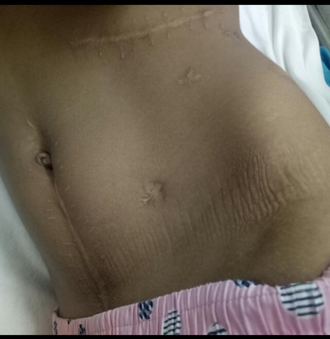
SITES DEMONSTRATED AS SURGICAL SCARS
4. PRE - OP. IVU X- RAY FROM 2019.
IVU X- RAY FROM 2019.
IT WAS INITIALLY REPORTED AS A DISTAL OBSTRUCTION IN THE DISCHARGE SUMMARY, WHICH IS DATED SEPTEMBER 28, 2019 (DISCHARGE DATE: 28-09-2019).
HOWEVER A CLOSER EXAMINATION SUGGEST'S THAT THE OBSTRUCTION IS NEAR PUJ???, WHICH MAKES IT VERY CLEAR THAT IT'S NOT A DISTAL OBSTRUCTION
FURTHER EVALUATION REQUIRED.
Date of discharge : 28/09/2019
Then the patient was discharged on 28/09/2019 with the following ADVICE:
Normal diet
TAB.SPORIDEX 125mg TID FOR 15 DAYS
SYP. PCM 250mg 2ml SOS
SYP. OSTEOCALCIUM 2ml OD FOR 15 DAYS
SYP. ZINCOVIT 5ml OD FOR 15 DAYS
SYP. TONOFEREL 4ml OD FOR 15 DAYS
N/K/C/O HTN; DM; TB; ASTHMA; EPILEPSY; CAD
INVESTIGATIONS IN SEQUENCE FROM 2019 TO PRESENT DAY
ULTRASOUND OF WHOLE ABDOMEN ON 08-05-2019
S.UREA and S.CREATININE ON 10-05-2019
URINE ANALYSIS ON 10-05-2019
CT SCAN KUB PLAIN and CONTRAST ON 13-05-2019
X RAY PART OF CHEST, ABDOMEN and PELVIS ON 23-05-2019
USG ON 23-05-2019
23-05-2019
RFT
LFT
24-05-2019
HBsAG RAPID and Anti HCV Antibodies RAPID
CBP ON 8-06-2019
SERUM ELECTROLYTES ON 19-06-2019
CBP ON 19-06-2019
CBP ON 9-7-19
RFT ON 9-7-19
USG - KUB REVIEW ON 22-07-2019
CBP ON 29-07-2019
BACTERIAL CULTURE and SENSITIVE REPORT ON 19-08-2019
CBP ON 21-08-2019
RFT ON 03-09-2019
CBP ON 03-09-2019

MICTURATING CYSTOURETHROGRAM ON. 7-6-19
CBP ON 14- 09-2019
ULTRASOUND OF WHOLE ABDOMEN ON 24-03-2023
EXAMINATION OF URINE ON 18-07-2023
USG ON 24-03-2024
S.CREARININE ON 1-4-2024
DTPA RENOGRAM ON 18-04-2024
URO REFERRAL on 30-04-2024
Impression: NO PUJ OBSTRUCTION
EMR SUMMARY
KIMS Hospital
Department: Urology
Patient
Age/Sex: 4 years / Male
Admission & Discharge
Admitted: 23/05/2019
Discharged: 26/10/2019
Discharge status: Relieved
Procedures Done
27/05/19: Nephrostomy
12/5/19: Posterior urethral valve fulguration
25/8/19: Left ureteric reimplantation and DJ stent insertion
17/9/19: Ureterostomy
Diagnosis
Left solitary kidney with PUV (Posterior Urethral Valve) with PUJ (Pelvi-Ureteric Junction) and left distal ureteric obstruction
Case History
Distension of left side of abdomen for 1 year
Polyuria
Increased urinary frequency
Condition at Discharge
“Condition uneventful at discharge” (stable)
Medications / Advice
Normal diet
Multiple medications for 15 days (including Sporinex, PCM, Cetirizine, Duphalac, Osteocalcium, Zincovit, Tonoferyl, etc.)
Avoid self-medication, don’t miss medications
Contact emergency / consultant doctor immediately in case of emergency
Follow-up
Review to Urology OPD after 15 days
Signatories(Treating Faculty)
(Senior Resident, Urology)
. (Junior Resident, Hospital Administration)
(Junior Resident, General Surgery)
(Faculty sign at discharge page)
Ultrasonography of whole abdomen of the Patient
Age: 6–7 years (reports from 2022–2023)
Sex: Male
Key Findings from Ultrasound Reports
Liver, Gall Bladder, CBD, Portal Vein, Pancreas, Spleen
All reports consistently show these organs to be normal in size, shape, echotexture and without focal lesions.
Gall bladder well distended, wall thickness normal, no calculi.
CBD and portal vein normal in calibre.
Pancreas and spleen normal.
Kidneys
Right Kidney: Not visualized / empty right renal fossa (suggests absent or ectopic right kidney).
Left Kidney:
Shows hydronephrosis (dilation of the kidney pelvis).
2022 report: Grade II hydronephrosis with cortical thinning and dilated renal pelvis, likely PUJ (pelvi-ureteric junction) obstruction. Size ~117.5 x 69.9 mm.
2023 report: Grade I hydronephrosis with mild cortical thinning and prominent renal pelvis. Size ~109.8 mm.
Impression: Left kidney is functioning but has some obstruction at PUJ level.
Urinary Bladder
Well distended.
Wall thickness mostly normal, but 2023 report mentions mild thickening (?cystitis).
Post-void residual urine: 31.2 cc (some urine left after urination).
Ureters & Prostate
Ureters: Not dilated.
Prostate: Normal for age.
Impressions Across Reports
Right kidney absent / not visualized (empty right renal fossa).
Left kidney with hydronephrosis and cortical thinning, likely due to PUJ obstruction.
Bladder wall mildly thickened (? cystitis).
-void residual urine present.
Advice for further evaluation: IVP / CT urogram, Urine RE/ME to rule out cystitis, and in some notes, CECT to rule out crossed kidney or other congenital anomalies.
Overall Summary
Child appears to have a single functioning left kidney with mild-to-moderate hydronephrosis (improving from grade II to grade I over time).
Right kidney not seen – could be absent (agenesis), ectopic, or fused.
PUJ obstruction suspected at left kidney.
Urinary bladder wall mild thickening with some residual urine suggests possible mild bladder dysfunction or infection.
Doctors are suggesting further imaging (CT/IVP) to clarify anatomy and congenital anomalies.


No comments:
Post a Comment